Picture of the Month: Living Cell Factories From the Institutes of Solid Mechanics and Biochemical Engineering
At first glance it could be the graphics of a thermal imaging camera. But in fact our picture of the month April shows a living cell factory under the microscope.The image was created in the course of a research cooperation between the Institute of Solid Mechanics and the Institute of Biochemical Engineering at the Technische Universität Braunschweig.
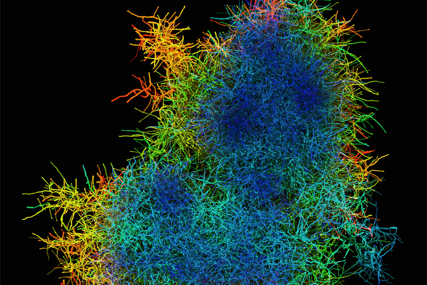
Depth-coded image of a filamentous biopellet of Actinomadura namibiensis (diameter approx. 150 µm). Picture credits: Markus Böl/TU Braunschweig
Filamentous microorganisms such as fungi or bacteria of the genus Actinomycetes are used and cultivated in biotechnology as living cell factories, for example to produce valuable enzymes, organic acids or pharmaceutical agents. Depending on the conditions under which these microorganisms are cultivated, they develop different forms (morphologies). Their filamentous cells (hyphae) can grow as mycelia, in which each hyphe is surrounded by the culture medium, or they can form dense, pellet-like structures in which the hyphal agglomerates combine to form a mostly spherical pellet.
The picture of the month shows the structure of such a pellet. Using a confocal microscope, a special light microscope, the scientists from the Institutes of Solid Mechanics and Biochemical Engineering of the TU Braunschweig have created a picture of a living cell factory. They have visualised the hyphal structure of a biopellet of the Actinomycetes bacterium Actinomadura namibiensis. The colour coding provides information on the position of the hyphae: blue hyphae are located close to the microscope objective, red ones are further away.
Compression of filamentous pellets
A characteristic of filamentous microorganisms is that the viability and productivity of their hyphae are closely linked to the morphology of the pellet and especially to the density of the hyphae. Their morphology is significantly influenced by changes in cultivation conditions, such as the addition of salt. This is shown, for example, by the fact that the addition of salt also significantly changes the pellet stiffness. The researchers from the Institutes of Solid Mechanics and Biochemical Engineering carried out compression experiments using a micromanipulator. This is a measuring device with which forces and paths in the micronewton or micrometer range can be recorded on individual biopellets. Biopellets were clamped between two plates and the force response exerted by the hyphal structure was determined over the distance between the plates. The results of these investigations were recently published in the Biochemical Engineering Journal.

Micromechanical compression experiment on a single pellet (diameter approx. 100 µm). The deformation increases from left to right. Picture Credits: Markus Böl/TU Braunschweig
Actinomadura namibiensis
As part of its metabolism, the bacterium A. namibiensis produces the antiviral agents Labyrinthopeptin A1 and A2. Labyrinthopeptin A1 shows antiviral activity against the Human Immunodeficiency Virus (HIV) and the Herpes Simplex Virus (HSV). In addition, synergistic effects with other standard antiretroviral drugs were found, which makes Labyrinthopeptin A1 a promising candidate for the development of a new broad-spectrum antibiotic. In contrast, Labyrinthopeptin A2 shows activity against neuropathic pain.