Picture of the Month April: Not one Cell like the Other Submitted by Professor Christian Sieben
They look like lots of colourful Easter eggs: the epithelial cells under super-resolution fluorescence microscopy. The picture of the month April shows an epithelial cell culture infected with influenza A viruses. Influenza A is considered the most dangerous type of influenza virus, responsible for severe pandemics and epidemics with many deaths. The picture was taken by Professor Sieben, head of the Nanoscale Infection Biology research group at the Helmholtz Centre for Infection Research and junior professor for cell biology of viral infections at the Technische Universität Braunschweig. The group uses special microscopy techniques to learn more about how viruses work and function, and thus how infections can be prevented.
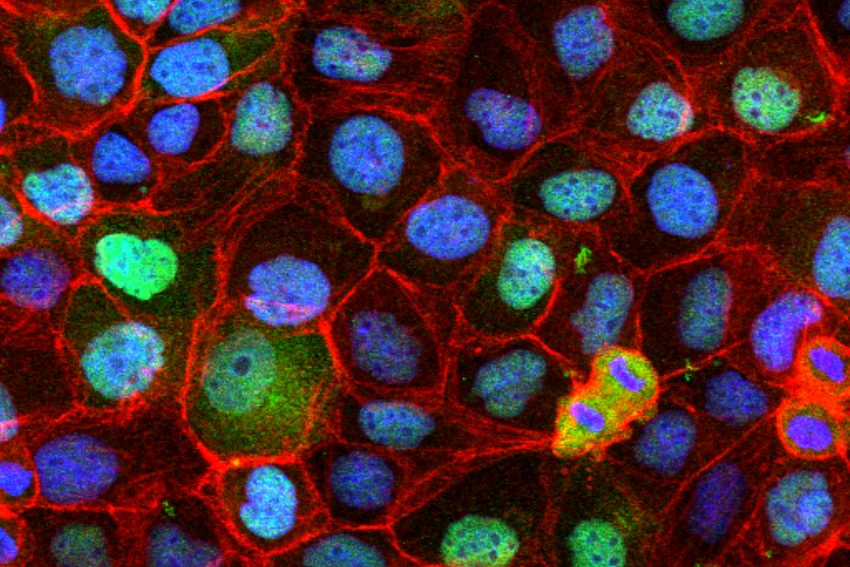
Epithelial cells under super-resolution fluorescence microscopy. Picture credit: Christian Sieben/TU Braunschweig
Epithelial cells can grow together to form a dense two-dimensional epithelium in which all cells seem to look identical. However, fluorescence microscopy shows that no two cells are alike. Influenza A viruses infect alveolar epithelial cells, so-called pneumocytes, among others in the lungs. The picture shows human A549 cells, a model cell line for human pneumocytes of type 2. The cells were infected with influenza A viruses and chemically fixated after about eight hours. To then detect the infection, the cells were treated with an antibody that binds the viral nucleoprotein (green).
The nucleoprotein is an important component of the influenza genome, which in turn is amplified in the cell nucleus. This leads to a strong accumulation of the nucleoprotein in the cell nucleus. In order to recognise the cell nucleus itself under the microscope, the DNA is marked with the dye DAPI (blue). In addition, the actin cytoskeleton, the protein network of thin, thread-like cell structures, is marked with a dye to identify the cell boundaries (red).
Each cell on its own
In this experiment, which is shown in the picture, it was examined how much the development of the infection differs from cell to cell. Although all cells were infected with approximately the same amount of virus, very strong differences can be seen between the cells.
“We are working on quantifying these effects and identifying their causes so that we can understand why individual cells seem to cope better with infection,” says Professor Sieben. To do this, his research group analyses the images on the computer. The labelling of the cellular structures (DNA and actin) enables the scientists to better identify the cells or the cell nucleus and thus precisely determine the amount of nucleoprotein produced per cell and even per cell nucleus.