Picture of the month: Microstructure of Skeletal Muscle – The Key to Understanding Our Body’s Movements From the Institute of Mechanics and Adaptronics
Healthy muscle activity is vital, not only for movement, but for many important processes such as speaking, chewing and breathing. In diseases associated with muscle atrophy, the microstructure of skeletal muscles is significantly altered. The Institute of Mechanics and Adaptronics at Technische Universität Braunschweig is working on the experimental multiscale characterisation of skeletal muscles, which can be used, for example, to make predictions about possible muscular deficits in patients in order to initiate appropriate treatment measures. Multiscale modelling also makes it possible to avoid animal experiments.
Skeletal muscles belong to the so-called active tissues that contract through an electrochemomechanical process and can thus generate active forces that lead to movements of our body. Crucial to this property is the special microstructure of skeletal muscles: A hierarchical, tubular architecture from the individual protein to the muscle fibre to the entire muscle. Disease-related changes in this finely tuned microstructure inevitably lead to an impairment of force generation and thus of the movement process.
The Institute of Mechanics and Adaptronics is involved in various research projects with experimental multiscale characterisation – from the individual muscle fibre to the muscle tissue to the entire muscle – as well as multiscale modelling of muscle tissue. Depending on the condition of the muscle, the microstructure varies greatly and depends, among other things, on age, gender and training condition. With the help of immunohistological techniques, the microstructure of skeletal muscles was made optically visible at the Institute of Mechanics and Adaptronics.
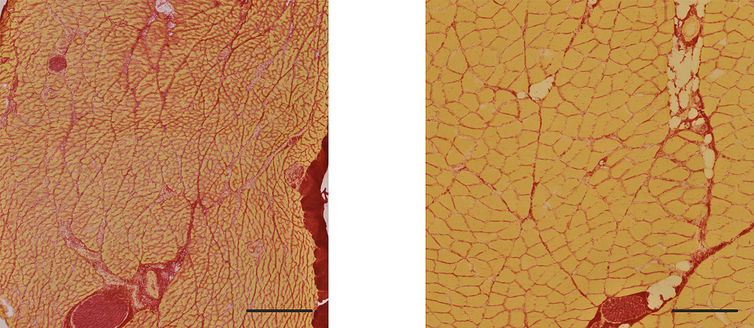
Typical histological muscle tissue sections – here of a rabbit – depending on age (age: 28 (left) and 87.5 days (right), prepared with Picro-Sirius red stain, collagen is stained red and muscle fibres are stained yellow. Scale: 300 μm. Photo credit: S.Kutschke/TU Braunschweig
Muscle cells are yellow, the structural protein collagen is coloured red. It can be clearly seen that muscle tissue shows marked differences in microstructure depending on age, which is of course also reflected in the active and passive load transfer mechanisms.
Furthermore, the multiscale modelling of such structures is of enormous importance: the numerical models offer the possibility to make predictions about the tissue without having to realise animal experiments. It is possible to incorporate tissue-specific or patient-specific information, such as mechanical properties at the fibre and collagen level, into the model.
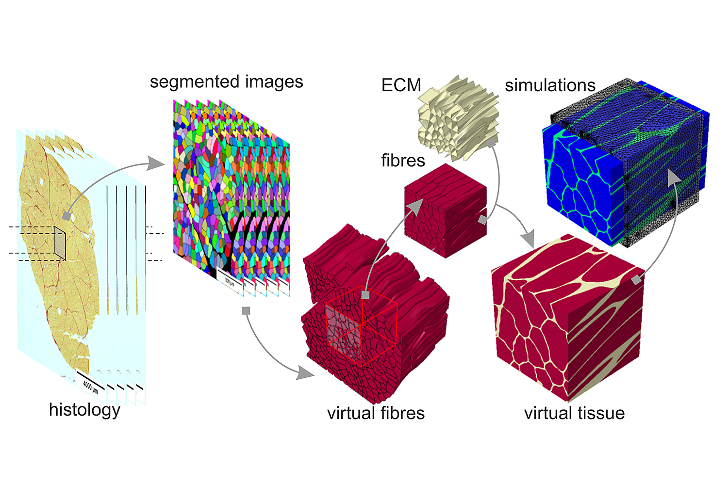
Procedure from histological sections to segmentation algorithms and virtual tissue to final simulation. Photo credit: K. Leichsenring/TUBS und A.E. Ehret, R. Kuravi/ETH Zürich